Skip Navigation

Log In
  • Scientific Research
  • Training
  • Professional Development
  • Diversity
  • Advocacy and Outreach
  • Career Paths
  • Image of three blue squares stacked vertically to look like pages. Collections
  • Advice For Annual Meeting
  • Think Alike?: A Neuronline Podcast
  • image of an open book Read
  • image of a play button: a triangle inside a circle Watch
  • an image of a calendar with a check mark signifying events to attend Attend
  • image of a blue microphone Listen
  • About Neuronline
  • SfN Events Calendar
  • Community Leaders Program
  • FAQ
  • Write for Us
  • Contact Us
Neuronline logo
SfN's home for learning and discussion
  • image of an open bookRead
  • image of a play button: a triangle inside a circleWatch
  • an image of a calendar with a check mark signifying events to attendAttend
  • image of a blue microphone Listen
Log In
  • Scientific Research
  • Training
  • Professional Development
  • Diversity
  • Advocacy and Outreach
  • Career Paths
  • COLLECTIONS

Scientific Research

Stay apprised of findings from across the field with summaries of research published in SfN journals, and learn about rigor, responsible conduct of research, and other topics important to the scientific community.
Thumbnail image for Getting Into Peer Review webinar
Professional Development
Getting Into Peer Review
July 10, 2025 12:00 PM - 1:00 PM EDT
Alzhiemer’s Relevance in the Neuroscience Community
Scientific Research
Alzheimer’s Relevance in the Neuroscience Community: A Collaborative Neuronline Perspective
Filter

Refine by

192 - 204 of 591
  • A woman stares intently at a computer.
    Ways to Improve Scientific Rigor While Working Remotely
    May 08, 2020
  • Enrichment map visualization for subcortical memory. Clusters are labeled with P for positive, N for negative.
    Distinct Genetic Signatures of Human Cortical and Subcortical Memory
    May 07, 2020
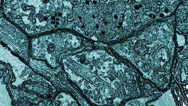
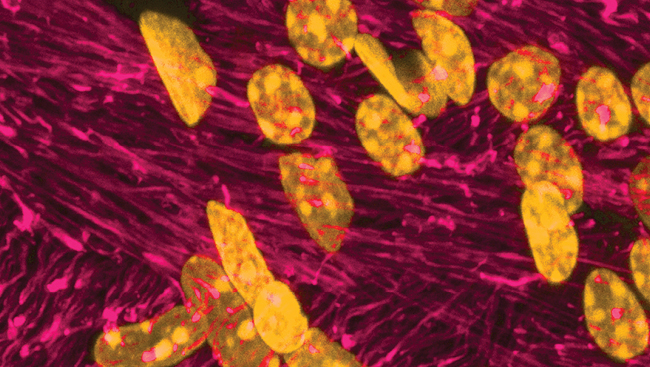
  • Cortex glia in the Drosophila larval nerve cord expressing GFP form a network surrounding neuronal cell bodies. Neuronal nuclei are stained orange and glial nuclei are stained blue.
    100 Years of Microglia
    Apr 21, 2020
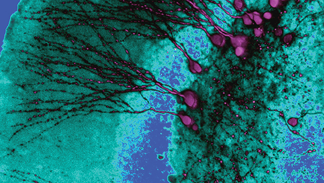
  • An image from the eNeuro research article "A New Mouse Line Reporting the Translation of Brain-Derived Neurotrophic Factor Using Green Fluorescent Protein."
    Monitoring Endogenous Growth Factor Translation in the Mouse Brain
    Apr 09, 2020
  • Shift of spatial representation from near Te in the target-aligned fixed window analysis toward Ge in saccade-aligned fixed window analysis of reactive task data. Each row shows the raster/spike density plot and best fit RF for an example neuron, followed the distribution of T-G α values of full population.
    Transforming Vision Into Brainstem Commands for Gaze Direction
    Mar 26, 2020
  • Predicting Future Diagnosis from Brain Data Alone
    Mar 25, 2020
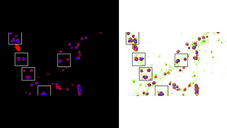
  • Image of sets of data collected and analyzed by Emily Hill.
    Tracking Tau Oligomer-Induced Pathology in Single Neuronal Cells
    Mar 12, 2020
  • A panelist speaks at a podium at a professional development workshop at Neuroscience 2019.
    Teaching Computation in Neuroscience
    Mar 05, 2020
  • Seven panelists sit at a table discussing social issues at Neuroscience 2019
    Ethical and Social Issues Raised by Neural-Digital Interfaces
    Mar 05, 2020
  • Optogenetic stimulation of the RE/CB reliably produces an evoked potential in the HPC.
    The Thalamic Nucleus Reuniens Orchestrates Prefrontal-Hippocampal Slow-Waves
    Feb 20, 2020
  • Movement trajectories are idiosyncratic between subjects, yet consistent within subject. Layout of the start position (green filled circle), and short (blue filled circle) and long targets (red filled circle) is the same as in Figure 1C. Color-coding of traces reflects hand movement through the progression of the given trial (black, warm-up period; green, during the tone duration; red, after the tone until the decision). Top row, Trajectory data for 2000 ms trials for three example subjects from Experiment 1 (free-movement). Classification of a short duration is the left target; a long classification is the right target. Each subject employed a separate strategy, with one subject (S4) moving in a circular pattern between short and long locations, another (S1) rotating in a leftward arc before moving in between both targets, and a third (S2) moving in an up–down fashion before shifting from the middle to a target location. Bottom row, Three example subjects for 2000 ms trials from Experiment 2 (hold). Changes-of-mind are evident, including a shift from long to short (S1), short to long (S3), and both (S2).
    Motor and Action Planning Enhance How We Make Decisions About Time Duration
    Feb 06, 2020
  • This image shoes the relative KCC2 depletion in WT and CX3CR1 BDNF KO animals 14 d after cut/ligation. Genotype had no effect on KCC2 levels in motoneurons contralateral or ipsilateral to injury. Removing BDNF from microglia had no impact on KCC2 expression. Lumbar spinal cord sections from animals expressing normal CSF1 and with CSF1 removed from motoneurons 14 d after ligation. ChatIREScre/+: : csf1f/f animals exhibit normal microgliosis in the dorsal horn but have the microglial reaction to injury greatly attenuated in the ventral horn compared to ChatI+/+: : csf1f/f. KCC2 immunofluorescence 14 d after sciatic ligation in csf1f/f animals. Preventing microgliosis in the ventral horn had no impact on KCC2 within intact or injured motoneurons. Error bars = SEM; **p ≤ 0.01, ***p ≤ 0.001.
    KCC2 on Axotomized Motoneurons Is Regulated Differently Than in Systems Previously Described
    Jan 23, 2020
  • Previous
  • 16
  • 17
  • 18
  • 19
  • 20
  • Next
Neuronline Collections
Neuronline's vast collection of professional development and training resources offers guidance for people at all career stages with diverse interests and responsibilities.
View Collections
Neuronline footer 10 year anniversary logo
  • About Neuronline
  • SfN Events Calendar
  • FAQ
  • Contact Us
  • Write for Us
  • Accessibility Policy
  • Disclaimer
  • Privacy Notice
SfN logo with "SfN" in a blue box next to Society for Neuroscience in red text and the SfN tag line that reads "Advancing the understanding of the brain and nervous system"
Follow SfN
  • BlueSky logo
  • Threads logo
  • X Logo
  • image of linkedin logo
  • Image of the Facebook logo
  • Image of the instagram logo
  • image of youtube logo
  • RSS symbol
1121 14th Street NW, Suite 1010, Washington, DC 20005 (202) 962-4000 | 1-888-985-9246

Copyright © 2025 Society for Neuroscience