Scientific Research
Stay apprised of findings from across the field with summaries of research published in SfN journals, and learn about rigor, responsible conduct of research, and other topics important to the scientific community.

Filter
Refine by
36 - 48 of 628
-
 April 22, 2026 1:00 PM - 2:00 PM EDT
 -
 August 20, 2025 12:00 PM - 1:00 PM EDT
 -
 September 16, 2025 12:00 PM - 1:00 PM EDT
 -
 July 10, 2025 12:00 PM - 1:00 PM EDT
 -
 June 24, 2025 12:00 PM - 1:00 PM EDT
 -
 Apr 14, 2025
 -
 May 14, 2025 11:30 AM - 12:30 PM EDT
 -
 April 22, 2025 12:00 PM - 1:00 PM EDT
 -
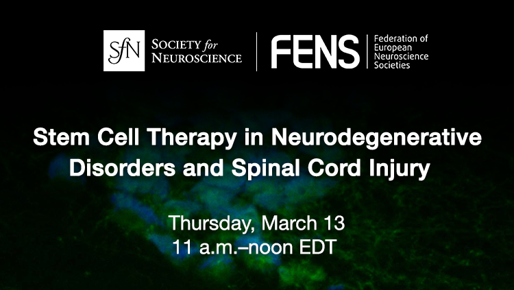 March 13, 2025 11:00 AM - 12:00 PM EDT
.png?h=423&w=750&la=en&hash=4890EC561E6BC83F646E0AD45CC1E31D666B7BEB)
.png?h=423&w=750&la=en&hash=F1FD544DD9CF144A59ACE1AEC3F87F3558711B1B)
.png?h=423&w=750&la=en&hash=2B26B20BD584E19DEC05CA1C07AFA8306ED2F98F)
Neuronline's vast collection of professional development and training resources offers guidance for people at all career stages with diverse interests and responsibilities.

