Skip Navigation

Log In
  • Scientific Research
  • Training
  • Professional Development
  • Diversity
  • Advocacy
  • Outreach
  • Career Paths
  • Image of three blue squares stacked vertically to look like pages. Collections
  • image of an open book Read
  • image of a play button: a triangle inside a circle Watch
  • an image of a calendar with a check mark signifying events to attend Attend
  • image of a blue microphone Listen
  • Image of two overlapping dialogue bubbles. Discuss
  • About Neuronline
  • Neuronline Leadership
  • FAQ
  • Write for Us
  • Contact Us
  • Community Guidelines
Neuronline logo
SfN's home for learning and discussion
  • image of an open bookRead
  • image of a play button: a triangle inside a circleWatch
  • an image of a calendar with a check mark signifying events to attendAttend
  • image of a blue microphone Listen
  • Image of two overlapping dialogue bubbles.Discuss
Log In
  • Scientific Research
  • Training
  • Professional Development
  • Diversity
  • Advocacy
  • Outreach
  • Career Paths
  • COLLECTIONS
    • All Collections
    • Advice for SfN's Annual Meeting
    • Best Practices for Mentoring Relationships
    • Foundations of Rigorous Neuroscience Research
    • Increasing Women in Neuroscience
    • Monthly Research Roundups
    • Optogenetics Training Series
    • Promoting Awareness and Knowledge to Enhance Scientific Rigor in Neuroscience
    • SfN Annual Meeting Recordings
    • SfN Journals: Research Article Summaries
    • Stem Cells and Reprogramming Methods for Neuroscience: An SfN Training Series
    • The Neuroscientist’s Guide to Advocacy

Scientific Research

Stay apprised of findings from across the field with summaries of research published in SfN journals, and learn about rigor, responsible conduct of research, and other topics important to the scientific community.
This image shows a parvalbuminexpressing inhibitory basket cell (green) in the hippocampus of an amyloid-depositing 5xFAD mouse. The cell has been morphologically reconstructed after patch clamp recording of spontaneous synaptic activity during sharp wave ripple events. Non-patched parvalbumin neurons are labeled in red.
Scientific Research
The Human Brain Tracks Speech More Closely in Time Than Other Sounds
Scientific Image
Scientific Research
Human Sensory Neurons Are Powerful Tools for Testing New Pain Treatments
Filter

Refine by

24 - 36 of 470
  • Extracting spatial heterogeneity of AMPA receptor content within functional zones of the synapses to quantify homeostatic scaling. Related to Figure 5 in the manuscript.
    New Immunostaining Analysis Reveals Spatial and Molecular Variability in Synaptic Scaling
  • Scientific Image
    Development and Regeneration: Common Themes and Important Differences
    September 09, 2020 11:00 AM - 4:00 PM EDT
  • Presynaptic infusion of G85R-SOD1 inhibited synaptic vesicle (SV) availability. Representative EM images illustrate morphology of AZs (labeled with red *) and numbers of SVs in fixed synapses infused with WT-SOD1 and G85R-SOD1.
    Synaptic Actions of an ALS-Associated Protein in the Giant Squid Synapse
  • The journey of cortical interneurons: (A) Cortical interneurons are born in the ganglionic eminence of the basal forebrain, and (B) migrate to reach the dorsal cortex. (C) Their number is determined upon programmed cell death and strongly depends on neuronal activity. (D) Wiring [dendritic and axonal morphogenesis and synapse establishment (1)], myelination (2) and circuit refinement of Martinotti, Basket, Chandelier and all other interneuron types is established within the first month after birth.  This cartoon has been designed and drawn by Yannis Maragkos, Denaxa Lab, BSRC Al. Fleming.
    How Cortical Interneurons Develop: Current and Future Research
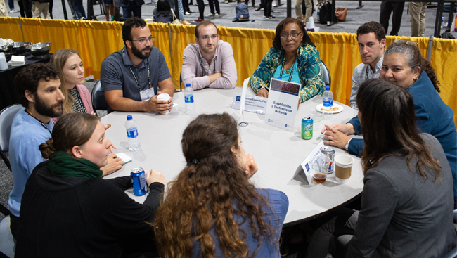
  • Female and male neuroscientists share ideas at a networking event.
    A Look at the Intersection of Scientific Rigor, Big Data, and Interdisciplinary Training
  • Scientific Image
    COVID-19, the Brain, and Mental Health: Weighing the Direct and Indirect Effects
    July 29, 2020 11:00 AM - 12:00 PM EDT
  • Images of graphs showing sex differences in cortical and dopaminergic neurodegeneration in 6-month-old 3K mice.
    Estrogen Benefits α-Synuclein Tetramerization and Motor Deficit in PD-Like Mice
  • Lesion histology.
    Superior Dorsolateral Prefrontal Cortex Supports Introspection in Monkeys
  • Scientific Image
    A Neurobiological Perspective on Emerging Mental Illness Research
  • Schematic representation of the molecular events following QR2 downregulation. Upon reduced QR2 expression or activity, either naturally or experimentally, there is a reduction in cellular ROS.
    QR2 Modulates Insular Cortex Redox to Enable Novel Taste Memory Formation
  • An image displaying the experimental design.
    Hippocampal CA2 Organizes CA1 Gamma Oscillations During Investigation of Novel Stimuli
  • This image shows tyrosine hydroxylase-immunoreactive neurons (green) in substantia nigra pars compacta. These neurons coexpress the dopamine transporter (red). In contrast, less than half of tyrosine-hydroxylase-expressing neurons in the hypothalamus expressed dopamine transporter, and those that don't are required for the autoregulation of growth hormone secretion.
    Voltage-sensitive Dye Imaging and Chemogenetics
  • Previous
  • 1
  • 2
  • 3
  • 4
  • 5
  • Next
Neuronline Collections
Neuronline's vast collection of professional development and training resources offers guidance for people at all career stages with diverse interests and responsibilities.
View Collections
Neuronline logo with Neuronline written in blue and gold text. Below the word Neuronline reads "Learning,  Discussion, Neuroscience"
  • About Neuronline
  • Neuronline Leadership
  • FAQ
  • Contact Us
  • Write for Us
  • Community Guidelines
  • Accessibility Policy
  • Disclaimer
  • Privacy Policy
SfN 50th Anniversary logo stating "Society for Neuroscience, celebrating 50 years. 1969-2019"
Follow SfN
  • image of linkedin logo
  • image of twitter logo
  • Image of the Facebook logo
  • Image of the instagram logo
  • RSS symbol
  • image of youtube logo
1121 14th Street NW, Suite 1010, Washington, DC 20005 (202) 692-4000

Copyright © 2019 Society for Neuroscience