Microscopy
Explore resources and articles on using microscopy techniques for neural circuit interrogation.
-
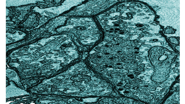 Bringing Two-Photon Microscopy Into Monkeys
Material below is adapted from the SfN Short Course Two-Photon Calcium Imaging in the Macaque Monkey, by Kristina J. Nielsen, PhD. Short Courses are daylong scientific trainings on emerging neuroscience topics and research techniques held the day before SfN’s annual meeting.
To learn more about the human brain, researchers often turn to looking at the brains of other primates, like the Rhesus macaque. Similar to the human brain in both organization and function, non-human primate brains can also be evaluated during tasks requiring complex thought, perception, and decision making. Understanding the neural circuitry underlying these processes will provide clues into the human brain, both its normal functioning and disruptions that lead to mental disorders. However, probing these circuits requires advanced techniques.
-
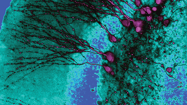 Shining a Light (Microscope) Onto the Brain's Connections
Material below is adapted from the SfN Short Course Mapping Brain Circuitry with a Light Microscope, by Pavel Osten MD, PhD and Troy W. Margrie, PhD. Short Courses are daylong scientific trainings on emerging neuroscience topics and research techniques held the day before SfN’s annual meeting.
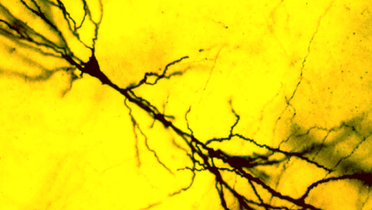
Since the days of Camillo Golgi and Santiago Ramón Y Cajal, light microscopes have provided neuroscientists a glimpse into the anatomy of the brain. Advances in the microscopes themselves and methods to stain and trace neurons have revealed various pathways and structures, as well as organization and hierarchies specific to certain brain regions. Three large projects (the Mouse Brain Architecture Project, the Allen Mouse Brain Connectivity Atlas, and the Mouse Connectome Project) are currently attempting to take our knowledge to another level, by mapping all the connections within the brain. Together with new technologies, these projects are helping bring the possibility of a complete map of the mouse brain within reach.
-
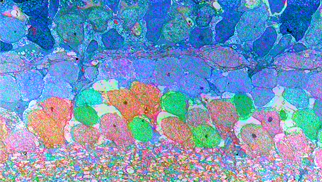 Widening the View of Two-Photon Microscopy
Material below is adapted from the SfN Short Course Mesoscale Two-Photon Microscopy Engineering a Wide Field of View with Cellular Resolution, by Jeffrey N. Stirman, PhD, and Spencer L. Smith, PhD. Short Courses are daylong scientific trainings on emerging neuroscience topics and research techniques held the day before SfN’s annual meeting.
Mapping activity in the brain requires both a wide lens — to see connections extending across brain regions — and a fine focus — to see the cells forming those connections. Two-photon microscopy coupled with calcium imaging meets the criteria for a fine focus; it can detect activity within individual neurons or within a local population of neurons. But beyond a small window, two-photon excitation lessens and the resolution suffers. This narrows the field of view, making mapping long-range connections impossible. Fortunately, newly described tweaks to both the microscopes and imaging techniques are making “mesoscale” two-photon microscopy possible, allowing wider views than can span brain regions a millimeter or more apart. Recently, this type of imaging has been performed in vivo to map activity in the visual system of mice.
-
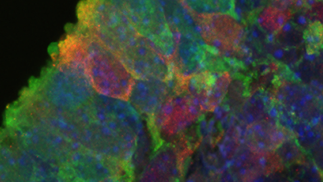 Seeing Neuronal Activity Simultaneously in Three Dimensions
Material below is adapted from the SfN Short Course Simultaneous Holographic Imaging of Neuronal Circuits in Three Dimensions, by Sean Quirin, PhD, et al. Short Courses are daylong scientific trainings on emerging neuroscience topics and research techniques held the day before SfN’s annual meeting.
Traditional microscopy techniques are limiting in that most only reflect two dimensions of a three-dimensional biological system. Optical imaging, such as two-photon microscopy to measure voltage-sensitive or calcium-sensitive dyes, is minimally invasive and has the resolution to study the individual cells that make up large neuronal networks. However, images from these experiments are captured in two dimensions; to collect data spanning a chunk of tissue, sequential images are scanned one at a time, losing any activity that occurs at the same time in different planes.
-
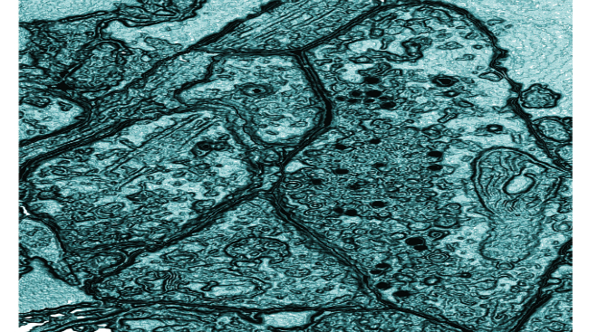
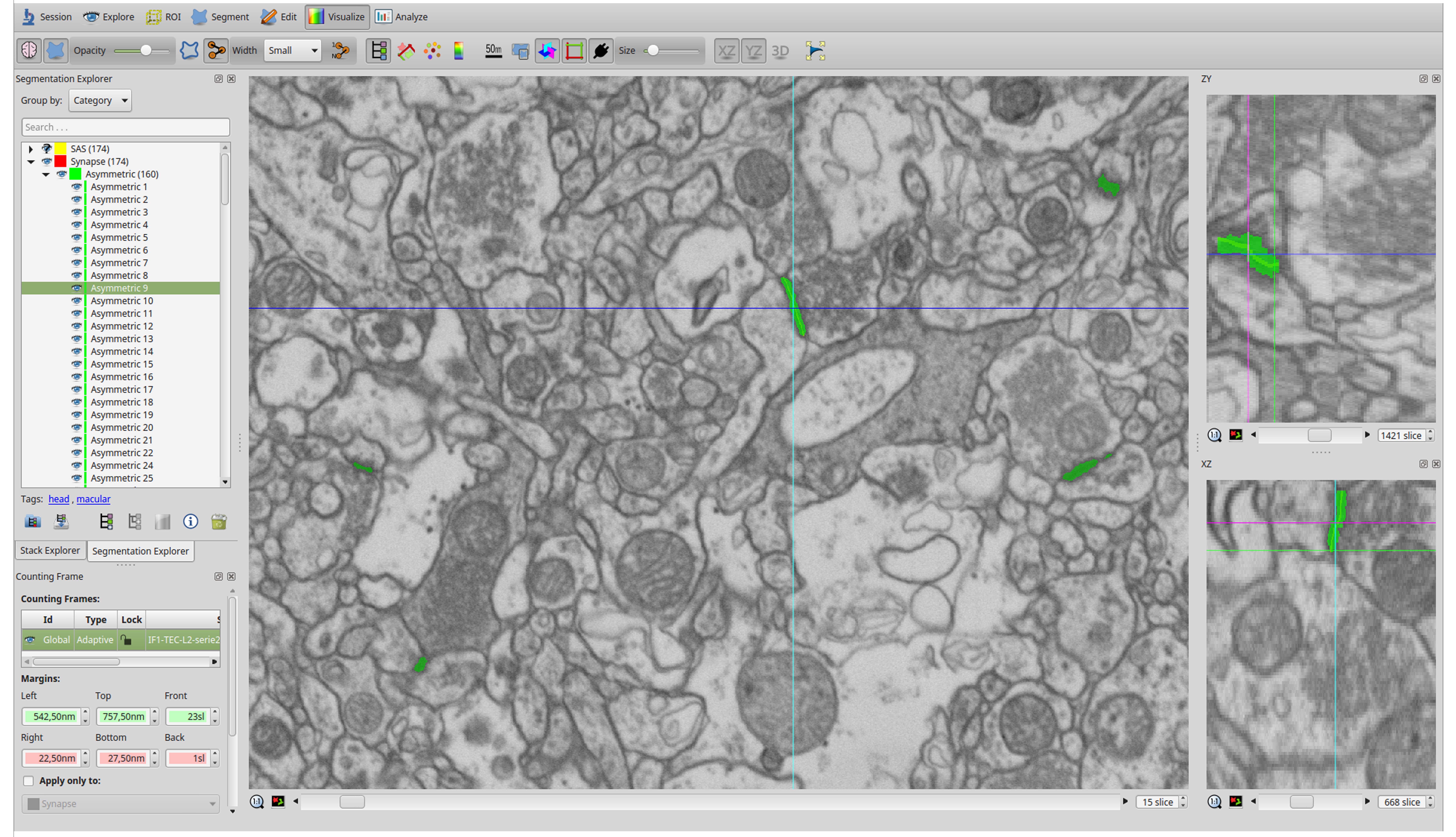
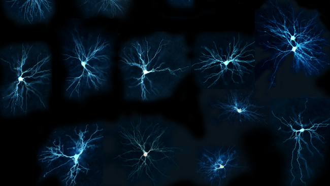 Synapses in the Human Transentorhinal Cortex
Material below summarizes the article 3D Electron Microscopy Study of Synaptic Organization of the Normal Human Transentorhinal Cortex and Its Possible Alterations in Alzheimer’s Disease, published on June 19, 2019, in eNeuro and authored by M. Domínguez-Álvaro, M. Montero-Crespo, L. Blazquez-Llorca, J. DeFelipe, and L. Alonso-Nanclares.
Highlights
- The majority of synapses are excitatory on spine heads (55%) and have a small disk shape. Nevertheless, a relatively large proportion of excitatory synapses are established on dendritic shafts (37%).
- Excitatory and inhibitory synapses show different preferences for their postsynaptic targets (spines or dendritic shafts).
- In Alzheimer’s disease (AD), morphological synaptic changes and a reduction in the number of synapses targeting spine heads occur.
-
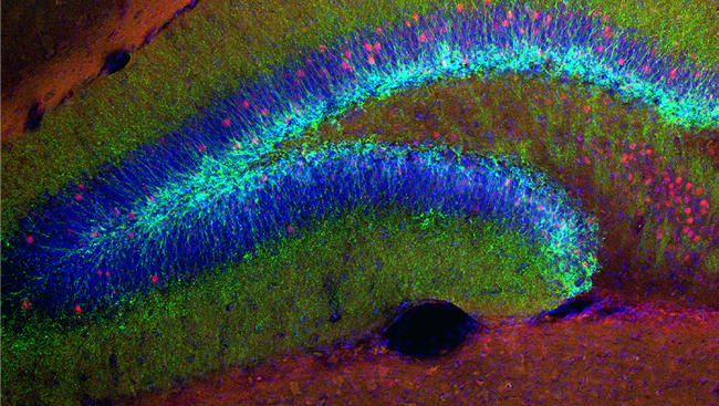 Studying Spinal Cord Axons with Better Transparency
Material below summarizes the article 3D Imaging of Axons in Transparent Spinal Cords from Rodents and Nonhuman Primates, published on March 26, 2015, in eNeuro and authored by Cynthia Soderblom, Do-Hun Lee, Abdul Dawood, Melissa Carballosa, Andrea Jimena Santamaria, Francisco D. Benavides, Stanislava Jergova, Robert M. Grumbles, Christine K. Thomas, Kevin K. Park, James David Guest, Vance P. Lemmon, Jae K. Lee, and Pantelis Tsoulfas.
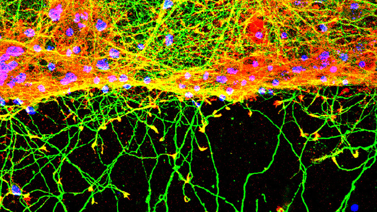
The beginning of this century has seen some major advances in light microscopy, particularly related to neuroscience. These developments in microscopy, coupled with techniques that make tissues transparent, are enabling microscopes to visualize the cellular architecture of whole tissues in 3D with unprecedented detail.
-
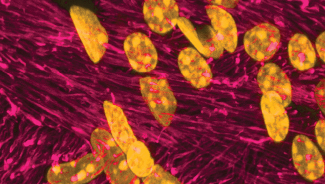 Fast, Focused, and Far-Reaching: A New 3D, Two-Photon Imaging Technique
Material below is adapted from the SfN Short Course Acousto-optical Scanning–Based High-Speed 3D Two-Photon Imaging in Vivo, by Balázs Rózsa, MD, PhD, Gergely Szalay, and Gergely Katona. Short Courses are daylong scientific trainings on emerging neuroscience topics and research techniques held the day before SfN’s annual meeting.
Oftentimes when imaging the brain, a wider view is sacrificed for finer detail, or vice versa. Although the ideal imaging technique could resolve both signals propagating within single neurons and signals sent across a whole network of hundreds of neurons, the two views present different challenges. Imaging in single cells must be able to distinguish activity in adjacent compartments (the soma compared to the dendrite, for example) while imaging networks of hundreds of cells must be able to measure simultaneous activity in cells that are hundreds of micrometers apart. Because of this, most current technologies are optimized for just one of the two views. A microscope that could tackle both would need to be fast yet fine-detailed, focused yet far-reaching.
-
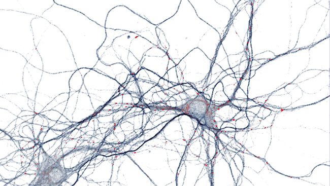 Microscopy Journal Articles
Read the latest research on the use of microscopy in neural circuit interrogation research.

