
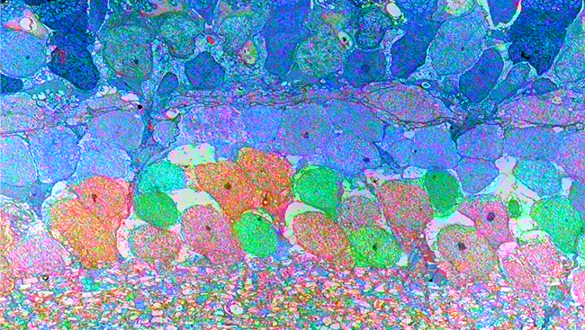
Material below is adapted from the SfN Short Course Mesoscale Two-Photon Microscopy Engineering a Wide Field of View with Cellular Resolution, by Jeffrey N. Stirman, PhD, and Spencer L. Smith, PhD. Short Courses are daylong scientific trainings on emerging neuroscience topics and research techniques held the day before SfN’s annual meeting.
Mapping activity in the brain requires both a wide lens — to see connections extending across brain regions — and a fine focus — to see the cells forming those connections. Two-photon microscopy coupled with calcium imaging meets the criteria for a fine focus; it can detect activity within individual neurons or within a local population of neurons. But beyond a small window, two-photon excitation lessens and the resolution suffers. This narrows the field of view, making mapping long-range connections impossible. Fortunately, newly described tweaks to both the microscopes and imaging techniques are making “mesoscale” two-photon microscopy possible, allowing wider views than can span brain regions a millimeter or more apart. Recently, this type of imaging has been performed in vivo to map activity in the visual system of mice.
Access to the full article is available to SfN members.
Neuronline is a benefit of SfN membership. Renew your membership now to make sure you don’t lose access.