Persistent Adaptations in Afferents to Ventral Tegmental Dopamine Neurons After Opiate Withdrawal
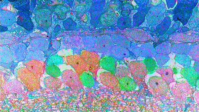
Material below summarizes the article Persistent Adaptations in Afferents to Ventral Tegmental Dopamine Neurons after Opiate Withdrawal, on July 15, 2015, in JNeurosci and authored by Jennifer Kaufling and Gary Aston-Jones.
The ventral tegmental (VTA) is a complex and heterogeneous structure. Dopaminergic (DA) neurons are a major population of VTA neurons and play a crucial role in reward processes, motivated behavior and drug addiction. Opiate addiction is a growing public health problem, and understanding the mechanisms of opiate actions in brain is crucial in order to treat dependent patients. For those reasons, we focused this study on the acute and chronic effects of morphine on the activity of VTA DA neurons.
A common characteristic of the acute effect of drugs of abuse is to directly or indirectly increase the activity of VTA DA neurons. Later, chronic use induces adaptive changes in the reward circuit that lead to addiction and related symptoms such as tolerance and withdrawal. Previous studies found that acute morphine substantially increases the basal activity of VTA DA neurons, and this increase is maintained during chronic morphine exposure but returns to control levels after acute or protracted withdrawal. Importantly, acute morphine fails to increase the basal activity of VTA DA neurons after protracted withdrawal, indicating that VTA DA neurons express morphine tolerance.
The regulation of VTA DA neuronal activity is complex and regulated by both inhibitory and excitatory inputs. VTA DA neurons are tonically inhibited by a resting GABA input that originates from several structures. One of these is a pool of GABAergic neurons just posterior to the VTA, named the tail of the VTA (tVTA) (also known as the rostromedial tegmental area). tVTA GABA neurons express the mu opiate receptor and project strongly to VTA DA neurons. In naïve animals, morphine inhibits tVTA neurons directly via these mu receptors, and this decreases tVTA inhibition of VTA DA neurons to indirectly activate (disinhibit) VTA DA neurons.
In view of those observations, we expected that chronic morphine exposure and morphine withdrawal would have opposite (mirror) effects on tVTA GABA neurons versus VTA DA neurons. Intriguingly, that was not the case.
Chronic morphine decreased the basal activity of tVTA GABA neurons, and withdrawal normalized this effect but only in the short term. Whereas the basal activity of VTA DA neurons had returned to naïve baseline after two weeks of morphine withdrawal, the basal activity of tVTA GABA neurons was lower in two week- withdrawn animals than in naïve animals. In addition, acute morphine still decreased tVTA GABA neuronal basal activity after two weeks of withdrawal — thus illustrating that tVTA GABA neurons do not develop morphine tolerance — in contrast to VTA DA activity, which was unaffected by acute morphine at this point.
These different adaptations to morphine led us to hypothesize a “disconnection” between tVTA GABA and VTA DA neurons during protracted withdrawal. However, we observed that direct stimulation of tVTA neurons inhibited VTA DA neurons both in naïve rats and after protracted withdrawal, whereas inhibition of tVTA cells failed to activate (disinhibit) DA neurons in animals after protracted withdrawal. This indicates that inhibitory signalling between tVTA and VTA remains functional but significantly altered during withdrawal. These results led us to hypothesize that the lack of activation of VTA DA neurons after opiate-induced inhibition of tVTA neurons during protracted withdrawal involves an additional factor.
As stated above, VTA DA neuronal activity is regulated by a complex balance of inhibitory and excitatory inputs, and we found that during protracted withdrawal both sides of this balance are disturbed. Thus, we showed that, in addition to decreased tVTA GABA activity, the resting level of baseline glutamatergic (excitatory) input to VTA DA neurons is greatly reduced in animals two weeks after morphine withdrawal. Therefore, the inhibition of tVTA neurons by acute morphine at this point cannot result in activation of VTA DA neurons because there is no underlying tonic excitatory drive on these DA neurons to activate them once the tVTA inhibition is removed.
Drug addiction is a chronic brain disorder, with deleterious consequences for individuals and their social environment, including the difficulty to remain abstinent after long-term withdrawal. Our data indicate that, after long-term opiate withdrawal, the tVTA remains able to inhibit VTA DA neurons but can no longer lead to their activation via disinhibition.
These results lead us to propose that the tVTA may maintain its capacity to transmit negative (aversive) information by inhibiting DA neurons, but can no longer permit the transmission of positive (rewarding) effects through disinhibition. This adaptation in the VTA DA reward system may contribute to negative symptoms linked with addiction, such as anxiety, depression, and anhedonia.
Visit JNeurosci to read the original article and explore other content. Read other summaries of JNeurosci and eNeuro papers in the Neuronline collection SfN Journals: Research Article Summaries.
Jennifer Kaufling and Gary Aston-Jones. (2015): Persistent Adaptations in Afferents to Ventral Tegmental Dopamine Neurons after Opiate Withdrawal, The Journal of Neuroscience, July 2015, 35(28): 10290-10303; DOI: 10.1523/JNEUROSCI.0715-15.2015