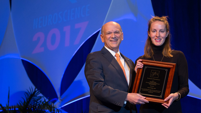
Katrin Franke’s research seeks to understand how the retinal network disassembles complex visual input. Previous research on this topic conducted by the field often focused on individual types of retinal cells, but Franke sought to record complete populations of neurons to capture the full functional diversity of parallel retinal channels. Through her approach, her findings have increased the understanding of how the mammalian retina processes visual information. For her outstanding work, she was awarded the Nemko Prize in Cellular or Molecular Neuroscience in 2017.
What led to your interest in visual processing?
Information processing in the visual system first attracted my interest as a master’s student at Tübingen University in a lecture series about the retina as a model system in neuroscience. I found it extremely fascinating that retinal cells perform computations to “decide” what’s important enough to be sent to the brain, determining what we see.
I decided to do a PhD in this field because I wanted to better understand how the retina decomposes the incoming visual stream into its relevant components that can then be interpreted by the brain.
In the last few years, I developed a strong interest in visual ecology, which aims to understand how different animal species use their visual systems to meet their ecological needs. I think investigating visual processing and comparing findings from different species is exciting and essential to discover universal and general principles of vision.
You developed a population approach to your particular research focus. What was your thinking behind taking this path?
The projects I worked on during my PhD sought to classify the neural cell types present in the inner retina of mice. In the last few decades, different approaches have been used to define neural cell types, many focusing on a single type of cell.
Alternately, my colleagues and I wanted to characterize a complete population of retinal cells in an unbiased way by combining large-scale functional imaging with unsupervised clustering. During our analysis, we found that combining information from different modalities of the same cells, like function and anatomy (in our case, light responses and soma size or stratification of axon terminals) critically improves the classification of cell types.
In my opinion, it will get increasingly important to work with multimodal data, including genetic, connectomic, and functional information. While this is already routinely done for single cells, extending this approach to neural populations will help to understand the function of different neural cell types in great detail.
What were some of the highs and lows you experienced when carrying out your experiments, including in the publishing process, for your thesis work, and how did you navigate through them?
Every PhD student goes through phases where nothing seems to be working without an obvious reason why. In these frustrating periods, I found it helpful to take a break for a few days and come back to the lab with a fresh mind.
Also, troubleshooting is much more efficient when done in a group rather than alone, which is why I always tried to seek the advice of other students in the lab. This helped me overcome most experimental problems without getting discouraged.
Regarding publishing, I really enjoyed the process of putting together a manuscript where all of the experimental data I acquired over a couple of years added up to an interesting story. In the end, it’s rewarding when people approach you to tell you they enjoyed reading your paper.
Currently, what questions are you interested in exploring, and why?
I’m still interested in visual information processing. There are still many exciting questions about how the neural processes in the early visual system relate to behavioral output.
In the future, I want to look beyond the retina and examine how the brain, and particularly the superior colliculus, makes sense of this information. In a new project, I would like to zoom into single visual features, such as color or motion, and follow them across multiple consecutive stages of the early visual system, including retina and higher visual centers in the brain.
I hope this approach will help us learn more about how visual information from photoreceptors is transformed into something “meaningful” for the brain as it travels through the different levels of the visual system.
Do you have any advice for other neuroscientists in the early stages of their careers?
I'd encourage young scientists to seek collaborations with colleagues from other disciplines, which is becoming increasingly important in science.
All of the studies I worked on during my PhD were highly collaborative, combining the expertise from different fields in neuroscience. Working on an excellent interdisciplinary team helped me broaden my scientific knowledge and analyze and interpret my data in a way that wouldn’t have otherwise been possible.
I’d also tell future PhD students to pay attention to the nonscientific attributes of a lab before they join.
I was lucky to work in a very supportive and inspiring environment that allowed me to develop my own research profile. In retrospect, this positive and relaxed atmosphere encouraged and strengthened my work.